Procedure
Percutaneous Pedicle Screw Fixation
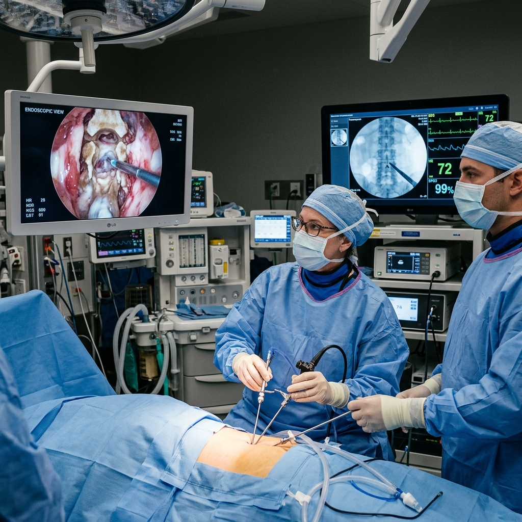
Percutaneous pedicle screw fixation achieves rigid spinal stabilisation through 18 mm incisions — one per screw — eliminating the open muscle stripping of conventional posterior spinal fixation.
About This Procedure
Pedicle screw fixation is the cornerstone of spinal stabilisation surgery. The percutaneous technique introduces screws through the pedicles — the strongest part of the vertebra — via small stab incisions under fluoroscopic or O-arm navigation, without exposing the spine. This preserves the posterior spinal musculature that is responsible for trunk extension strength and long-term functional outcomes. Rods are then passed through the screw tulips subcutaneously, creating a rigid construct. Percutaneous fixation is used for fractures, spondylolisthesis, fusion augmentation, and adjacent segment disease. Dr. Sparsh Jaiswal utilises both fluoroscopic and O-arm 3D navigation for percutaneous screw placement, achieving < 2% malposition rates.
Indications
- check_circleTraumatic lumbar fractures requiring stabilisation without laminectomy
- check_circleLumbar spondylolisthesis requiring posterior fixation
- check_circleSupplement to interbody fusion procedures (MIS-TLIF)
- check_circleSpinal tumour stabilisation after vertebrectomy
- check_circleAdjacent segment disease after prior fusion requiring extension
Step-by-Step Procedure
Image-Guided Planning
Fluoroscopic or O-arm CT imaging establishes the precise trajectory for each screw. Navigation arrays or anatomical landmarks guide entry points.
Percutaneous Entry
A Jamshidi needle is advanced through a 5 mm stab incision to the pedicle entry point under lateral and AP fluoroscopic guidance. Position confirmed.
Sequential Screw Insertion
A guide wire is placed, the pedicle is tapped, and the screw is advanced over the wire. The process is repeated for all planned levels (typically 4–6 screws).
Rod Passage & Final Tightening
A connecting rod is passed through the screw heads percutaneously and locked. Compression or distraction applied as required. Final fluoroscopic check confirms construct.
What to Expect
Surgery takes 60–90 minutes for a 2-level construct. Patients mobilise within 24 hours. Incisions are small (four to six 18 mm wounds). Mild back soreness from screw tracks settles in 1–2 weeks.
Recovery
Hospital stay is 2–3 days. Lumbar support for 4–6 weeks. Driving in 3–4 weeks. Return to desk work in 2–3 weeks; manual labour in 6–8 weeks. Hardware removal is rarely necessary.
Risks & Considerations
verified_userMedically reviewed: May 2026 — Dr. Sparsh Jaiswal, MBBS DNB FISS FINM MNAMS · Apollo Hospitals, Indore (Reg: MP-16286)
Frequently Asked Questions about Percutaneous Screw Fixation
What is Percutaneous Screw Fixation?
Pedicle screw fixation is the cornerstone of spinal stabilisation surgery. The percutaneous technique introduces screws through the pedicles — the strongest part of the vertebra — via small stab incisions under fluoroscopic or O-arm navigation, without exposing the spine. This preserves the posterior spinal musculature that is responsible for trunk extension strength and long-term functional outcomes. Rods are then passed through the screw tulips subcutaneously, creating a rigid construct. Percutaneous fixation is used for fractures, spondylolisthesis, fusion augmentation, and adjacent segment disease. Dr. Sparsh Jaiswal utilises both fluoroscopic and O-arm 3D navigation for percutaneous screw placement, achieving < 2% malposition rates.
What can I expect during Percutaneous Screw Fixation?
Surgery takes 60–90 minutes for a 2-level construct. Patients mobilise within 24 hours. Incisions are small (four to six 18 mm wounds). Mild back soreness from screw tracks settles in 1–2 weeks.
What is the recovery after Percutaneous Screw Fixation?
Hospital stay is 2–3 days. Lumbar support for 4–6 weeks. Driving in 3–4 weeks. Return to desk work in 2–3 weeks; manual labour in 6–8 weeks. Hardware removal is rarely necessary.
What are the risks of Percutaneous Screw Fixation?
Screw malposition (< 2% with navigation) — rarely symptomatic. Pedicle fracture during screw insertion (< 1%). Nerve root irritation from medially placed screw. Hardware failure (rod fracture) in long-level constructs. Wound haematoma or superficial infection (1–2%).
Other Procedures
Discuss this procedure with Dr. Sparsh
Book a consultation to find out if you are a suitable candidate and understand the full process in detail.