Procedure
Endoscopic Discectomy
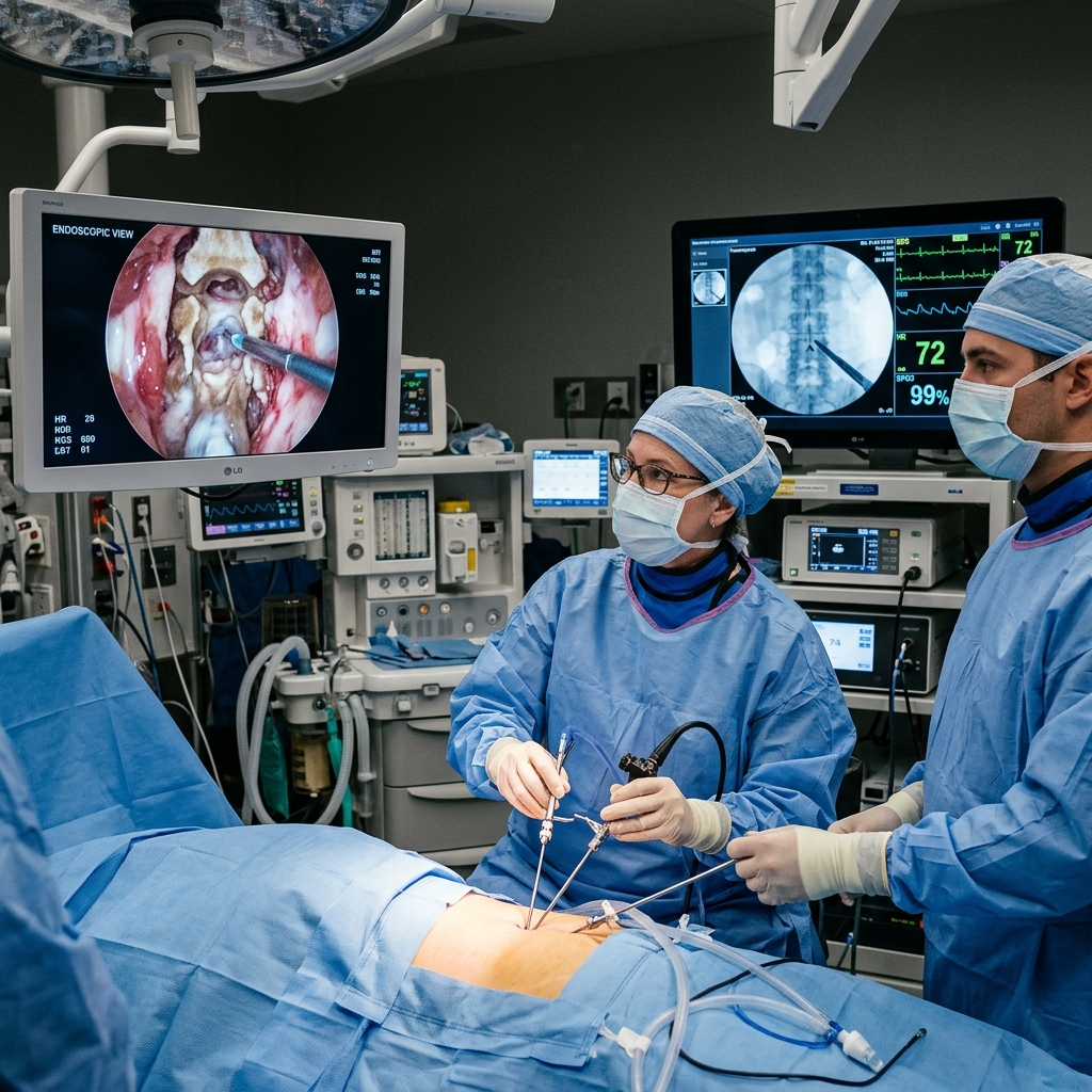
Endoscopic discectomy is the most minimally invasive surgical option for herniated disc treatment — performed through a 7 mm incision under direct camera visualisation, with patients walking within hours.
About This Procedure
Endoscopic discectomy removes the herniated disc fragment that is compressing a spinal nerve, through a single 7 mm working portal. The endoscope provides high-definition, magnified visualisation of the disc fragment, nerve root, and surrounding structures — enabling precise, safe removal with minimal trauma to surrounding tissues. Unlike open or microscopic discectomy, endoscopic discectomy can often be performed under epidural anaesthesia, allowing the conscious patient to provide real-time neurological feedback. Dr. Sparsh Jaiswal trained in this technique at IBS Hospitals, New Delhi, and offers both transforaminal and interlaminar approaches based on disc location.
Indications
- check_circleLumbar disc herniation causing sciatica unresponsive to 6 weeks of conservative management
- check_circleCervical disc herniation causing arm pain or radiculopathy
- check_circleFar-lateral disc herniation not accessible by standard tubular approach
- check_circleAcute severe neurological deficit from disc compression
- check_circleRecurrent disc herniation after previous open surgery
Step-by-Step Procedure
Positioning & Anaesthesia
Patient placed prone (lumbar) or supine (cervical) under epidural or general anaesthesia. Fluoroscopic localisation of the herniation level confirmed.
7 mm Portal Creation
A 7 mm incision is made. Sequential dilators create a working channel to the herniation. No muscle cutting — fibres are dilated apart.
Endoscopic Disc Removal
Under HD endoscopic visualisation with continuous saline irrigation, the herniated fragment is precisely removed using endoscopic forceps and laser. Nerve root decompression confirmed.
Haemostasis & Closure
Endoscopic bipolar ensures haemostasis. The working cannula is removed. A single absorbable suture or skin glue closes the 7 mm portal.
What to Expect
Operation time is 45–75 minutes. Most patients experience immediate relief of leg or arm pain. Walking is permitted within 2–4 hours. Mild post-operative back soreness resolves within days.
Recovery
Discharge same day or next morning. Driving in 5–7 days. Return to desk work in 3–7 days; physical labour in 3–4 weeks. No spinal brace required. Physiotherapy initiated at 2–3 weeks for core strengthening.
Risks & Considerations
Other Procedures
Discuss this procedure with Dr. Sparsh
Book a consultation to find out if you are a suitable candidate and understand the full process in detail.