Procedure
Endoscopic Spinal Fusion
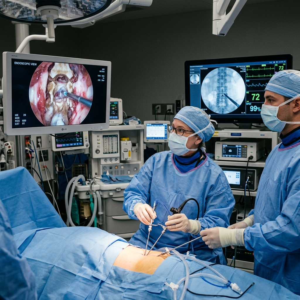
Endoscopic spinal fusion combines the decompressive accuracy of endoscopy with percutaneous instrumentation to achieve stable lumbar fusion through incisions measuring just 7–18 mm.
About This Procedure
Endoscopic transforaminal lumbar interbody fusion (Endo-TLIF) is an evolution of standard MIS-TLIF, performing the interbody portion entirely through a full-endoscopic portal before supplementing with percutaneous pedicle screws. This approach achieves complete disc space preparation, interbody cage insertion, and fusion without the muscle damage of conventional open fusion. It is particularly suited for degenerative spondylolisthesis, disc degeneration causing chronic pain, and isthmic spondylolisthesis. Dr. Sparsh Jaiswal applies this technique to achieve fusion results comparable to open surgery with the recovery profile of endoscopic decompression.
Indications
- check_circleDegenerative lumbar spondylolisthesis causing stenosis and instability
- check_circleIsthmic spondylolisthesis in active patients
- check_circleSevere discogenic back pain with Modic endplate changes after failed conservative treatment
- check_circleAdjacent segment disease after prior lumbar fusion
- check_circleLumbar disc herniation with concurrent instability
Step-by-Step Procedure
Percutaneous Screw Placement
Four percutaneous pedicle screws placed bilaterally under fluoroscopic guidance through 18 mm incisions — one per screw site.
Endoscopic Disc Preparation
Through a 7 mm endoscopic portal, the disc space is thoroughly cleared of nucleus material, and the endplates are prepared to bleeding bone for fusion.
Cage & Graft Insertion
An appropriately sized PEEK or titanium interbody cage packed with bone graft (autologous or synthetic) is inserted endoscopically to restore disc height and achieve fusion.
Rod Passage & Finalisation
Connecting rods are passed percutaneously through the screw heads and tightened under fluoroscopic confirmation. Final imaging confirms alignment and construct position.
What to Expect
Surgery takes 2–3 hours. Patients mobilise on post-operative day 1. Mild incision soreness resolves within 1–2 weeks. Fusion consolidation takes 3–6 months and is monitored with CT imaging.
Recovery
Hospital stay is 2–3 days. A lightweight lumbar support may be worn for 4–6 weeks. Return to desk work in 2–3 weeks; manual work in 8–12 weeks. Physiotherapy-based rehabilitation from week 3.
Risks & Considerations
Other Procedures
Discuss this procedure with Dr. Sparsh
Book a consultation to find out if you are a suitable candidate and understand the full process in detail.